About the Laboratory
Our laboratory studies how cells decide to divide, and how that decision goes wrong in cancer. Cell-cycle control cannot be understood from genotype alone, or from a single snapshot in time. In many settings it is the timing, strength, and duration of signaling, read out one cell at a time, that determines whether a cell divides, enters a reversible resting state, or, in cancer, becomes tolerant to a targeted drug. To study this, we combine genetically encoded live-cell biosensors, high-throughput quantitative microscopy, single-cell tracking and image analysis, optogenetics, CRISPR functional-genomic screens, and in vivo tumor models, which let us watch signaling and cell-cycle decisions unfold in individual cells over time. Our work moves between two connected questions: how the cell cycle is normally controlled, and how cancer cells rewire that control to escape therapy.
Watching persisters emerge in real time
MCF-7 breast cancer cells expressing a live-cell CDK2 activity sensor, imaged during treatment with the CDK4/6 inhibitor palbociclib. As most cells arrest, a subset escapes and reactivates CDK2, the drug-tolerant persisters that seed resistance. Single-cell imaging lets us catch this divergence as it happens, within 48 hours of treatment, rather than inferring it after the fact.
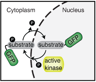
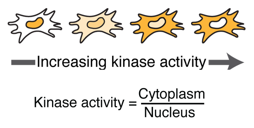
CDK2 kinase translocation reporter
Basic Cell-Cycle Regulation
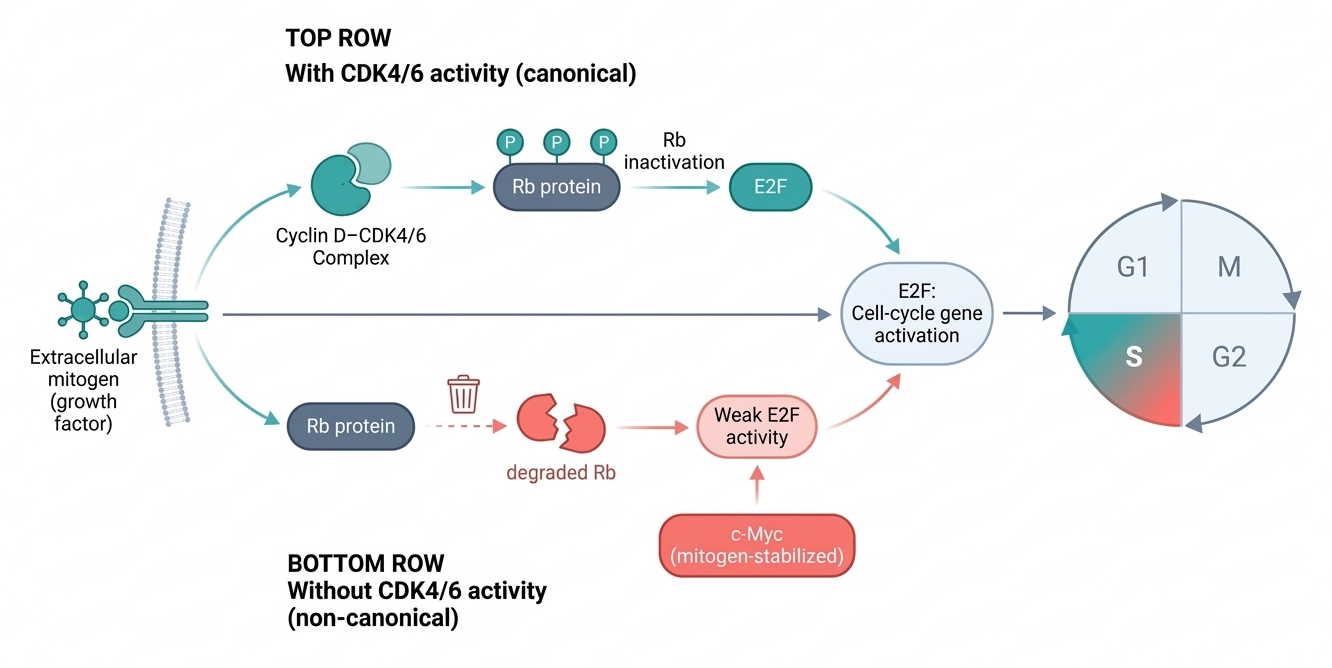
How does a cell decide to divide, and how is that decision kept under control? A core part of our program is the basic biology of cell-cycle regulation. Cells normally commit to division when the kinases CDK4 and CDK6 inactivate the retinoblastoma protein (Rb), releasing E2F transcription factors that switch on the genes for DNA replication and drive the G1-to-S transition. We study how this commitment is made, and we have found it is more flexible than the textbook switch suggests.
We discovered a non-canonical route to cell-cycle entry that operates even when CDK4/6 activity is absent: unphosphorylated Rb is intrinsically unstable, so its degradation can relieve E2F repression on its own. This route is only permissive, producing weak E2F activity, and robust proliferation requires a second input, mitogen-stabilized c-Myc, that amplifies E2F to the threshold for S-phase entry. This work showed that cells can bypass CDK4/6 dependence while still preserving tight control over when they divide.
We have extended this logic beyond G1. In G2-arrested cells, we identified a mitogen-dependent decision, governed by E2F activity, that determines whether a cell re-enters mitosis or instead exits the cycle and undergoes whole-genome duplication (WGD). Because WGD is a major route to chromosomal instability, oncogene amplification, and aggressive tumor behavior, we now study how cells sense and respond to genome doubling, how they choose between arrest and proliferation afterward, and how these early decisions shape cell fate and genomic instability over time. Together, this work treats cell-cycle entry not as a simple checkpoint, but as a dynamic, history-dependent process that integrates signaling, Rb stability, E2F activity, and growth cues at single-cell resolution.
Dysregulation of the Cell Cycle in Cancer
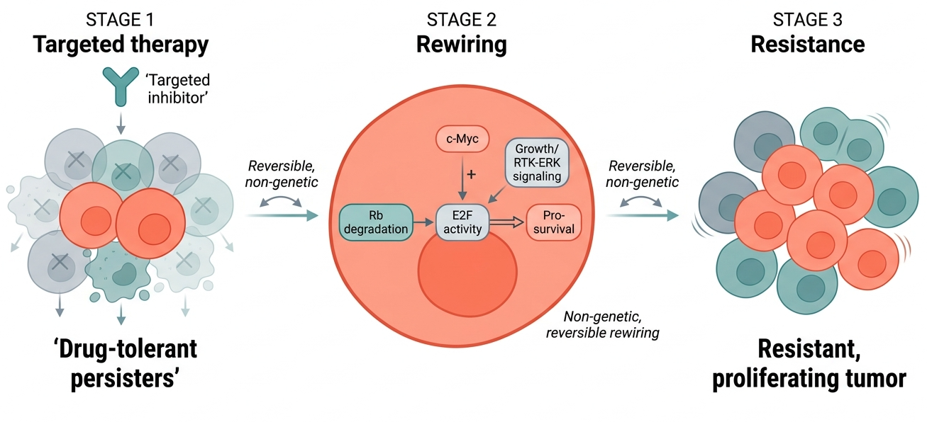
Cancer cells survive by rewiring the very pathways that normally control division. The same flexibility that lets a healthy cell tune its decision to divide becomes, in cancer, a route to escape therapies designed to stop proliferation. We study how tumor cells remodel cell-cycle and signaling circuitry to adapt to targeted inhibition, often without acquiring any new mutation, and how that adaptation can be prevented or reversed.
A recurring theme in our work is non-genetic adaptation. When a targeted drug suppresses proliferation, a small subset of cells does not die but instead enters a reversible, drug-tolerant persister state, and resistant tumors later regrow from these survivors. We have found that cancer cells reach this state by reactivating the same Rb, E2F, and c-Myc circuitry we study in normal cells: treatment relieves E2F repression, and growth signaling amplifies it back to a proliferative threshold. Because the rewiring is built on regulation rather than mutation, it is in principle reversible, which makes it a promising point of intervention.
We pursue this concept across cancer types and therapies. In breast cancer, we study how cells adapt to CDK4/6 inhibition and how combining it with CDK2 or CDK7 inhibition can suppress resistance and strengthen antitumor immunity. In melanoma, we study how the timing and strength of RTK and ERK signaling let a subset of cells rebound and re-enter the cycle after BRAF, MEK, or RAS inhibition. Across these settings, our goal is the same: to define the logic cancer cells use to rewire cell-cycle control, and to turn that understanding into strategies that make targeted therapy more durable.